
IPAK-EDU Director’s Science Webinar
Monday 3/16 @ 7:00pm Eastern
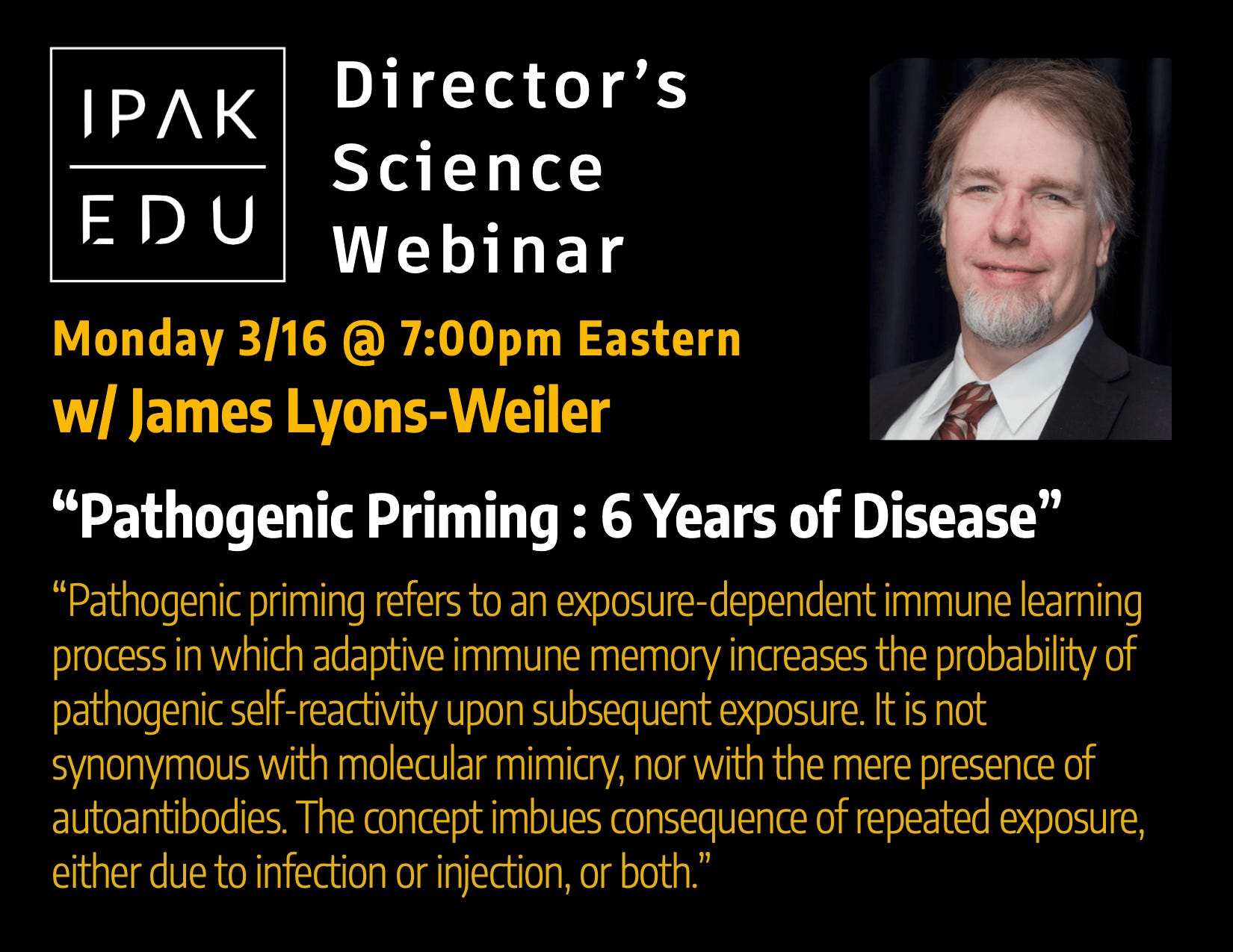
w/ James Lyons-Weiler, PhD
“Pathogenic Priming”
from the Popular Rationalism substack:
Pathogenic Priming Nearly Six Years Out: What Do We Know??
In April 2020, “Pathogenic priming likely contributes to serious and critical illness and mortality in COVID‑19 via autoimmunity” introduced a specific concept, backed by data, that repeated exposure to immunogenic viral epitopes that share homology with human proteins would likely prime the immune system toward pathogenic autoimmunity, with consequences that extend beyond acute infection to multi‑system disease and long‑term morbidity.
That paper, funded by the public via IPAK, did not merely assert that “molecular mimicry is possible.” It catalogued predicted autoreactive homology between SARS‑CoV‑2 epitopes and human proteins across immune‑relevant pathways, showed that only one immunogenic epitope lacked human homology, and explicitly warned that exposure by infection or injection carried foreseeable autoimmune risk if those homologous regions were used uncritically in antigen design.¹
Nearly six years later, the literature citing that work no longer sits at the level of conjecture. It contains experimental demonstrations of antibody cross‑reactivity, functional autoantibodies with physiological effects, validated biomarker panels that discriminate post‑acute sequelae of COVID‑19 (PASC), post‑vaccination prolonged‑symptom cohorts with defined autoantibody signatures, tissue‑level immune injury documented at autopsy, and population‑scale shifts in autoimmune disease incidence. The question in 2026 is no longer whether pathogenic priming is biologically plausible. The question is which parts of the causal chain have been empirically observed, which endpoints are now measurable, and where precision still fails.
This article synthesizes that record using the PubMed‑indexed citation corpus associated with the original 2020 paper, frozen as of January 2026, and focuses on what has been observed, not merely predicted.
Introduction: from warning to record
In April 2020, Pathogenic priming likely contributes to serious and critical illness and mortality in COVID‑19 via autoimmunity introduced a specific claim, not a rhetorical concern: repeated exposure to immunogenic viral epitopes that share homology with human proteins can prime the immune system toward pathogenic autoimmunity, with consequences that extend beyond acute infection to multi‑system disease and long‑term morbidity. That paper did not merely assert that “molecular mimicry is possible.” It catalogued homology between SARS‑CoV‑2 epitopes and human proteins across immune‑relevant pathways, showed that only one immunogenic epitope lacked human homology, and explicitly warned that exposure by infection or injection carried foreseeable autoimmune risk if those homologous regions were used uncritically in antigen design.¹
Nearly six years later, the literature citing that work no longer sits at the level of conjecture. It contains experimental demonstrations of antibody cross‑reactivity, functional autoantibodies with physiological effects, validated biomarker panels that discriminate post‑acute sequelae of COVID‑19 (PASC), post‑vaccination prolonged‑symptom cohorts with defined autoantibody signatures, tissue‑level immune injury documented at autopsy, and population‑scale shifts in autoimmune disease incidence. The question in 2026 is no longer whether pathogenic priming is biologically plausible. The question is which parts of the causal chain have been empirically observed, which endpoints are now measurable, and where precision still fails.
This article synthesizes that record using the PubMed‑indexed citation corpus associated with the original 2020 paper, frozen as of January 2026, and focuses on what has been observed, not merely predicted.
Defining pathogenic priming operationally
Pathogenic priming refers to an exposure‑dependent immune learning process in which adaptive immune memory increases the probability of pathogenic self‑reactivity upon subsequent exposure. It is not synonymous with molecular mimicry, nor with the mere presence of autoantibodies. It is defined by measurable phenomena that can be independently assessed:
Demonstrable cross‑reactivity between anti‑pathogen immune effectors and self‑antigens.
Functional immune effects (receptor activation, inhibition, complement fixation, immune complex formation).
Reproducible associations between immune markers and clinical phenotypes.
Organ pathology consistent with immune‑mediated injury.
Changes in disease incidence or diagnosis patterns at the population level.
The concept imbues consequence of repeated exposure, either due to infection or injection, or both.
The literature now occupies each of these tiers.
The 2020 starting point and its testable predictions
The 2020 analysis compared immunogenic SARS‑CoV‑2 epitopes across all viral proteins to the human proteome and found pervasive high‑local homology.¹ Mapping those human matches to pathways identified targets in MHC class I and II antigen presentation, PD‑1 signaling, cross‑presentation of soluble antigens, and ER‑phagosome pathways—precisely the systems that govern tolerance, exhaustion, and immune regulation. The translational implication was explicit: homologous epitope regions represent pathogenic priming risk and should be excluded or redesigned in vaccine development.
Two classes of predictions followed. First, that antibodies or T‑cell responses directed at SARS‑CoV‑2 would cross‑react with human proteins in susceptible individuals. Second, that such cross‑reactivity would manifest clinically as autoimmune or immune‑mediated disease, particularly with repeated exposure.
Evidence layer I: Epitope homology and immunoinformatic refinement
Post‑2020 work expanded epitope mapping using immunologically informed criteria. Moody et al. identified 136 alignments (6–23 amino acids) between SARS‑CoV‑2 proteins and 129 human proteins, emphasizing extracellular accessibility and immunogenic likelihood rather than raw identity counts.² Subsequent immunoinformatic screens narrowed these predictions to specific B‑cell‑recognizable regions and extended them to CD4⁺ T‑cell autoepitopes presented by defined HLA class II alleles.³ These studies moved the field from “there is homology” to “here are candidate epitopes, here are candidate host proteins, and here are the host genotypes most likely to present them.”
These analyses alone do not establish disease. Their importance lies in specifying where empirical validation should occur—and that validation did occur.
Evidence layer II: Experimental antibody cross‑reactivity
In 2021, Vojdani, Vojdani, and Kharrazian applied human monoclonal antibodies against SARS‑CoV‑2 spike and nucleoprotein, and polyclonal antibodies against envelope and membrane proteins, to a panel of 55 human tissue antigens.⁴ They observed immune reactivity with 28 of 55 antigens, spanning barrier proteins, gastrointestinal tissues, thyroid, and neural targets. Selective epitope mapping using BLAST demonstrated homology between viral proteins and human antigens including mitochondrial M2, F‑actin, and thyroid peroxidase.
This study is frequently dismissed by critics as “only binding.” That dismissal misses its significance. Monoclonal antibodies collapse the ambiguity of polyclonal serum noise. A monoclonal that binds a human antigen above threshold establishes specificity, not coincidence. The study did not claim clinical causation. It demonstrated a necessary mechanistic step: anti‑SARS‑CoV‑2 antibodies can bind self‑antigens relevant to observed disease phenotypes.
Evidence layer III: Functional autoantibodies with physiological effects
The transition from binding to function occurred decisively in Long COVID research. Wallukat et al. investigated 31 patients with persistent post‑COVID symptoms and detected functionally active autoantibodies (fAABs) targeting G‑protein‑coupled receptors in all 31.⁵ Each patient carried between two and seven receptor‑active autoantibodies acting as agonists or inverse agonists, measured using a neonatal rat cardiomyocyte bioassay as the functional readout.
The targets—β₂‑adrenergic, α₁‑adrenergic, AT1, muscarinic M2, MAS, ETA, and nociceptin receptors—map directly onto dysautonomia, tachycardia, fatigue, tremor, cognitive impairment, and cardiovascular instability, all hallmark Long COVID symptoms. This is not theoretical autoimmunity. It is receptor‑level immune interference with demonstrable physiological effects leading to disease.
Evidence layer IV: Biomarker‑level discrimination of PASC
Hatayama et al. advanced the field further by coupling discovery‑scale screening to validation‑grade diagnostics.⁶ Using a ~20,000‑protein bead array, they identified candidate autoantibodies associated with PASC, then validated two—PITX2 and FBXO2—by ELISA in expanded cohorts including PASC patients, non‑PASC convalescents, and pre‑pandemic controls. PITX2 autoantibodies discriminated PASC from non‑PASC convalescents with an AUC of 0.891 and from healthy controls with an AUC of 0.866. FBXO2 showed moderate but consistent performance.
Importantly, these autoantibodies correlated with specific symptoms, including dyspnea, palpitations, appetite loss, and cognitive impairment. This represents a critical maturation point: autoimmunity linked not only to exposure but to diagnosable, reproducible post‑acute disease phenotypes.
Evidence layer V: Post‑vaccination prolonged‑symptom cohorts
Mantovani et al. examined a retrospective case series of 17 individuals who were healthy before vaccination, reported no prior SARS‑CoV‑2 infection, and experienced prolonged PACVS‑like symptoms for a median of 20 months after vaccination.⁷ High proportions carried autoantibodies against GPCRs and renin–angiotensin system–related molecules. Specific autoantibodies associated with symptom clusters, including anti‑ACE2 with skin manifestations and anti‑MAS1 with widespread burning sensations. Anti‑ACE2 levels correlated with anti‑spike antibodies, consistent with an anti‑idiotype mechanism.
This study does not estimate incidence and does not disentangle all confounding. What it does establish is that prolonged post‑exposure syndromes with defined autoantibody signatures are not confined to infection‑only cohorts.
Evidence layer VI: Tissue‑level immune injury
Autopsy studies provide an independent line of evidence. Macor et al. examined lung, kidney, and liver tissue from 12 patients who died of COVID‑19 and demonstrated deposition of complement components C1q, C4, C3, and C5b‑9 on vascular endothelium, with IgG colocalization consistent with classical pathway activation.⁸ Complement deposition extended across organs, supporting systemic immune‑mediated injury rather than isolated viral cytopathy.
Tissue pathology does not identify epitopes. It does constrain mechanism: immune complexes, antibody‑mediated complement activation, and vascular injury are not speculative when visualized directly.
Evidence layer VII: Autoimmune diagnoses and syndromes
Case series and systematic reviews document new‑onset immune thrombocytopenia, vasculitis, reactive arthritis, necrotizing myopathy, dysautonomia, and other autoimmune conditions temporally associated with SARS‑CoV‑2 infection or vaccination.⁹⁻¹³ While individual case reports cannot establish causality alone, their consistency across systems and response to immunomodulatory therapy reinforce an immune‑mediated mechanism.
Notably, counter‑examples exist. Gasparini et al. found anti‑phospholipid antibodies in 34.7% of hospitalized COVID‑19 patients yet observed no association with thrombotic events in that cohort.¹⁴ This matters. It demonstrates that not all autoantibodies are pathogenic, and it sets the evidentiary bar correctly: functional relevance and endpoint linkage are required.
Evidence layer VIII: Population‑scale autoimmune incidence shifts
Sliman et al. analyzed data from over 1.5 million children in Israel, comparing autoimmune disease incidence before and after the pandemic onset.¹⁵ They reported significant increases in juvenile idiopathic arthritis, systemic lupus erythematosus, Henoch–Schönlein purpura, autoimmune thyroid disease, type 1 diabetes, psoriasis, vitiligo, and later immune thrombocytopenia, with heterogeneous patterns across gastrointestinal diseases. The authors explicitly noted diagnostic‑practice confounders for some conditions, strengthening rather than weakening the analysis by making biases explicit.
Population‑level shifts do not identify epitopes. They do establish that the autoimmune landscape changed in measurable ways following mass exposure to SARS‑CoV‑2 antigens.
Synthesis: what has been observed
Taken together, the literature now documents a partial but substantial causal chain:
Extensive epitope homology between SARS‑CoV‑2 and human proteins.
Experimental antibody cross‑reactivity to tissue antigens.
Functional autoantibodies with receptor‑level physiological effects.
Validated autoantibody biomarkers that discriminate PASC.
Prolonged post‑vaccination syndromes with defined autoantibody profiles.
Tissue‑level immune injury consistent with antibody‑mediated mechanisms.
Population‑scale shifts in autoimmune disease incidence.
What remains unresolved is not whether pathogenic priming occurs, but which epitopes, which host genotypes, and which exposure histories lead to specific endpoints. Most importantly, what therapies can be designed to alleviate or reverse the autoimmunity caused by exposure to unsafe epitopes, and how can vaccines be design that exclude unsafe epitopes?
Implications and remaining precision gaps
The central unresolved variable is repeated exposure. Few studies yet quantify dose–response relationships linking exposure count, epitope specificity, immune repertoire evolution, and clinical outcome within the same individuals. That gap is methodological, not conceptual.
The original 2020 warning regarding antigen design remains intact. Homologous epitope regions are identifiable. Functional consequences of immune targeting are now observable. Ignoring those facts in future vaccine or therapeutic development would be a choice, not an oversight.
Conclusion
Pathogenic priming is no longer a speculative construct. It is a framework supported by binding data, functional assays, biomarker validation, tissue pathology, clinical syndromes, and epidemiology. The task ahead is precision: mapping specific immune targets to specific outcomes under defined exposure conditions, treatments and prevention. The evidence base now supports that work. Pretending otherwise is no longer scientifically defensible.
References
Lyons‑Weiler J. Pathogenic priming likely contributes to serious and critical illness and mortality in COVID‑19 via autoimmunity. J Transl Autoimmun. 2020;3:100051. doi:10.1016/j.jtauto.2020.100051
Moody R, Wilson KL, Boer JC, et al. Predicted B‑cell epitopes highlight the potential for COVID‑19 to drive self‑reactive immunity. Front Bioinform. 2021;1:709533. doi:10.3389/fbinf.2021.709533
Timofeeva AM, Aulova KS, Mustaev EA, Nevinsky GA. SARS‑CoV‑2 spike protein and molecular mimicry: an immunoinformatic screen for cross‑reactive autoantigen candidates. Int J Mol Sci. 2025;26(18):8793. doi:10.3390/ijms26188793
Vojdani A, Vojdani E, Kharrazian D. Reaction of human monoclonal antibodies to SARS‑CoV‑2 proteins with tissue antigens. Front Immunol. 2021;11:617089. doi:10.3389/fimmu.2020.617089
Wallukat G, Hohberger B, Wenzel K, et al. Functional autoantibodies against G‑protein‑coupled receptors in patients with persistent Long‑COVID‑19 symptoms. J Transl Autoimmun. 2021;4:100100. doi:10.1016/j.jtauto.2021.100100
Hatayama Y, Miyakawa K, Kimura Y, et al. Identification of putative serum autoantibodies associated with post‑acute sequelae of COVID‑19. Int J Mol Sci. 2025;26(4):1751. doi:10.3390/ijms26041751
Mantovani M, Bellavite P, Fazio S, et al. Autoantibodies targeting GPCRs and RAS‑related molecules in post‑acute COVID vaccination syndrome. Biomedicines. 2024;12(12):2852. doi:10.3390/biomedicines12122852
Macor P, Durigutto P, Mangogna A, et al. Multiple‑organ complement deposition on vascular endothelium in COVID‑19 patients. Biomedicines. 2021;9(8):1003. doi:10.3390/biomedicines9081003
Bhattacharjee S, Banerjee M. Immune thrombocytopenia secondary to COVID‑19: a systematic review. SN Compr Clin Med. 2020;2:2048‑2058. doi:10.1007/s42399‑020‑00521‑8
Karampoor S, Afrashteh F, Rahmani S, Laali A. Eosinophilic granulomatosis with polyangiitis after COVID‑19. Respir Med Case Rep. 2022;38:101702. doi:10.1016/j.rmcr.2022.101702
Kocyigit BF, Akyol A. Reactive arthritis after COVID‑19: a case‑based review. Rheumatol Int. 2021;41:2031‑2039. doi:10.1007/s00296‑021‑04998‑x
Tsuzuki‑Wada T, Yokota K, Inayoshi F, et al. Immune‑mediated necrotizing myopathy after SARS‑CoV‑2 mRNA vaccination. Intern Med. 2023;62:3699‑3706. doi:10.2169/internalmedicine.2551‑23
Eldokla AM, Numan MT. Postural orthostatic tachycardia syndrome after mRNA COVID‑19 vaccine. Clin Auton Res. 2022;32:307‑311. doi:10.1007/s10286‑022‑00880‑3
Gasparini G, Canepa P, Verdiani S, et al. Anti‑phospholipid antibodies in hospitalized COVID‑19 patients. Int J Immunopathol Pharmacol. 2021;35:20587384211042115. doi:10.1177/20587384211042115
Sliman RKA, Cohen H, Shehadeh S, et al. Pediatric autoimmune diseases in the light of COVID‑19 pandemic. J Transl Autoimmun. 2025;10:100281. doi:10.1016/j.jtauto.2025.100281
Read more at Popular Rationalism.

Links from the archive
Once a week, this webinar series brings a focus to science as it applies to public issues of concern. The webinar brings to you expertise from scientists, researchers, data analysts, medical professionals, health advocates, attorneys, and more, all with the opportunity to learn, ask questions, and hear directly from people working at the leading edge.
Join us!

If you like what you see, and you’re willing and able, consider leaving a tip. Thank you!